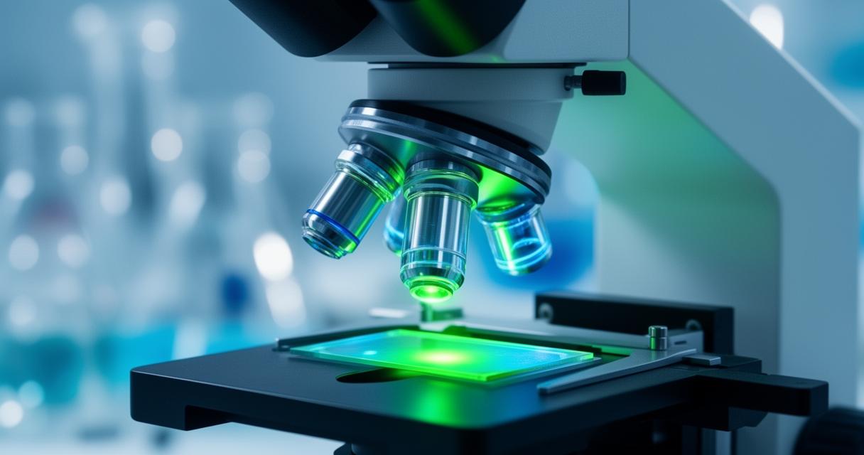
From the earliest handcrafted lenses to today’s sophisticated electron beams, microscopy has quietly shaped our understanding of the world at scales invisible to the naked eye. Whether examining biological tissue, testing new alloys, or inspecting semiconductor components, the ability to magnify and resolve fine detail underpins countless breakthroughs across science and industry. This article takes a closer look at how these tools work, the innovations driving them forward, and why they remain indispensable to researchers and manufacturers alike.
Key points
- Optical microscopy uses visible light for affordable and accessible imaging, whereas electron microscopy utilizes electron beams to achieve superior resolution for detailed material analysis.
- Resolution is the critical measure of a microscope’s effectiveness, defining the ability to distinguish fine details at the nanometer scale.
- Scanning Electron Microscopy (SEM) and Transmission Electron Microscopy (TEM) are essential for examining surface topography and internal structures in materials science and manufacturing.
- Fluorescence and phase contrast microscopy provide non-destructive ways to observe delicate biological samples and transparent living cells in real time.
- Historical developments, beginning with the pioneering work of Antoni van Leeuwenhoek, have evolved into modern cutting-edge technologies like Atomic Force Microscopy.
- Advanced imaging techniques are indispensable for quality control and research across diverse fields, including semiconductors, pharmaceuticals, and materials engineering.
Fundamental Principles and Types of Microscopy
Understanding optical and electron microscopy
At its core, microscopy is the practice of observing samples that are simply too small to be seen with the naked eye. The most familiar approach is optical microscopy, which relies on visible light and a series of glass lenses to magnify an image. These instruments remain popular in classrooms and laboratories because they are relatively affordable and straightforward to operate, making them ideal for basic research and educational resources alike. However, when researchers need to peer far deeper into the structure of a material, they often turn to electron microscopy. Rather than light, this technique fires a focused beam of electrons at a specimen, producing images with far greater detail than any conventional lens could achieve. This distinction matters enormously across fields such as materials science, semiconductors, and life science research, where the tiniest structural feature can hold the key to a discovery.
The Role of Resolution in Imaging Clarity
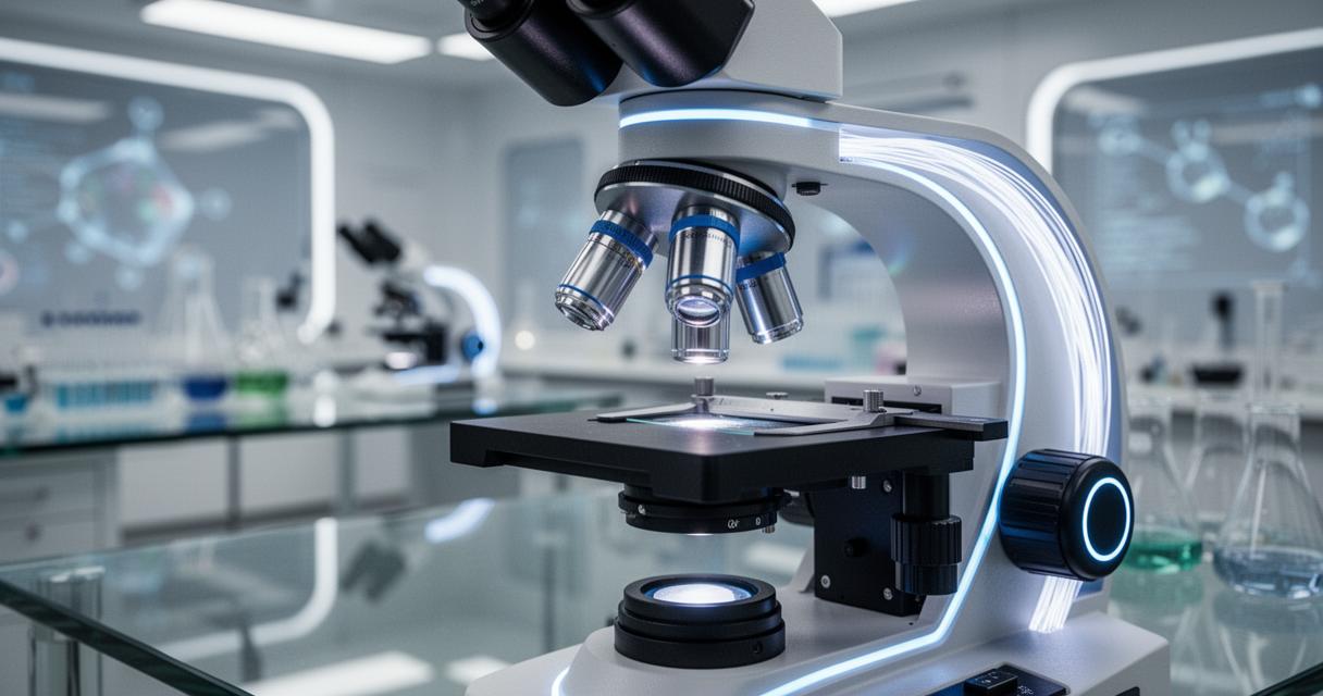
Resolution is, in many respects, the defining measure of any microscope’s usefulness. It refers to the ability to distinguish between two points that are extremely close together, and it dictates just how much genuine detail an image can reveal. Optical systems are inherently limited by the wavelength of visible light, whereas electron-based systems can achieve resolution many times finer, allowing scientists to examine features at the nanometre scale. This is precisely why industries ranging from pharma to quantum technologies depend on high-resolution imaging tools; without sharp, reliable clarity, subtle defects or structural quirks could easily be overlooked, with consequences for both research outcomes and product quality control.
Advanced microscopy methods for detailed analysis
Scanning and transmission electron microscopy techniques
Among the most widely used advanced methods is Scanning Electron Microscopy, commonly known as SEM, which scans a focused electron beam across the surface of a sample to build a detailed topographical image. This makes it especially useful for studying the texture and composition of materials such as metals, alloys, composites, and ceramics, and it has become a mainstay in advanced manufacturing and geology alike. A related but distinct technique, Transmission Electron Microscopy or TEM, instead passes electrons straight through an extremely thin sample, revealing internal structures that would otherwise remain hidden. Combining these approaches with focused ion beam technology, often referred to as FIB-SEM, allows researchers to slice through a sample layer by layer, producing three-dimensional insights that are invaluable in fields like forensics, biomaterials research, and semiconductor inspection. Suppliers such as ZEISS and Leica Microsystems have played a considerable role in refining these instruments, ensuring they remain accessible to laboratories working across chemical, catalysis, and energy storage research.
Fluorescence and phase contrast approaches
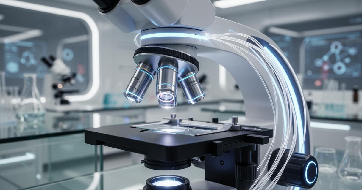
Not every specimen benefits from electron bombardment, particularly delicate biological samples that need to remain intact and, ideally, alive during observation. This is where fluorescence microscopy proves its worth, using fluorescent dyes or naturally glowing proteins to highlight specific cells or cellular components under particular wavelengths of light. It has become a cornerstone technique in bio imaging and clinical diagnostics, offering researchers a way to track processes in real time. Meanwhile, phase contrast microscopy addresses a different challenge altogether, that of observing transparent or colourless specimens that would otherwise be nearly impossible to see under a standard light microscope. By converting subtle differences in light phase into visible contrast, this method has become a go-to tool for studying living cells without the need for staining, a real advantage in basic research and drug discovery settings where preserving sample integrity matters.
Historical Milestones and Modern Innovations in Microscopy
Contributions of Antoni van Leeuwenhoek and Research Institutions
No discussion of microscopy would be complete without acknowledging Antoni van Leeuwenhoek, the Dutch tradesman whose handcrafted lenses in the seventeenth century allowed him to observe bacteria and other microorganisms for the very first time. His curiosity and craftsmanship effectively laid the groundwork for the entire discipline, inspiring generations of scientists to refine and expand upon his rudimentary designs. In the centuries since, institutions such as the Pasteur Institute have carried this legacy forward, using microscopy to make significant advances in medicine and microbiology. Today, that same spirit of enquiry continues at research centres and universities worldwide, many of which collaborate closely with instrument manufacturers to push the boundaries of what these tools can achieve.
Cutting-Edge Developments in Optics and Imaging Technology
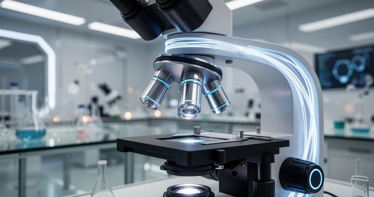
Modern microscopy has come a remarkably long way from Van Leeuwenhoek’s simple glass beads. Advances in electronics and optics have given rise to techniques such as laser scanning and tunnelling microscopy, both of which allow researchers to probe surfaces and structures with astonishing precision. Atomic Force Microscopy, for instance, uses a tiny mechanical probe rather than light or electrons to map surfaces at an atomic scale, proving invaluable in photonics, polymers, and nanotechnology research. It is worth noting that several Nobel Prizes in Chemistry have been awarded for breakthroughs directly tied to microscopy, underscoring just how transformative these innovations have been across scientific disciplines. Platforms like SelectScience now bring together news, application guides, and webinars covering everything from spectroscopy to lab automation, helping practitioners stay informed as the field continues to evolve. Looking ahead, the fusion of digital microscopes with automated analysis and improved sample preparation methods promises to make these tools even more accessible, whether the application lies in astronomy, environmental studies, or the ongoing pursuit of new therapeutic applications in medicine.